LA Orthopedic Group can help diagnose and treat Tarsal Coalition.
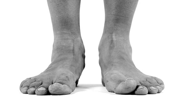
Tarsal coalition is a type of flat foot that occurs when more than two bones in the midfoot or hindfoot (tarsal bones) join together. The tarsal bones include talus, calcaneus, cuboid, navicular and cuneiform bones. These bones work in harmony to provide the motion needed for normal foot function.
- Most are born with the condition
- Symptoms typically manifest when bones start to mature, between the ages of 10 and 16
CONTACT US TODAY
Types of Tarsal Coalition
The two most common types of tarsal coalitions include:
- Calcaneonavicular coalition – involves the navicular bone and the calcaneus bone.
- Talocalcaneal coalition – involves the calcaneus bone and the talus bone.
Other forms of tarsal coalition include calcaneocuboid coalition, talonavicular coalition and cubonavicular coalition.
Symptoms of Tarsal Coalition
In some instances, the symptoms are not present in childhood but develop later in life. Tarsal coalition symptoms may include:
- Pain when walking or standing
- Fatigue and tired or cramped legs
- Flatfoot (in one or both feet)
- Muscle spasms in the leg
- Stiffness of the foot and ankle


Causes of Tarsal Coalition
Tarsal coalition is an inherited condition, meaning that it runs in families. Most often, the condition occurs when individual bones in a developing embryo fail to separate properly. Tarsal coalition can develop in later life as a result of infections, arthritis, and a previous injury to the area.
How it’s Diagnosed
Diagnosis of tarsal coalition includes obtaining information about the age and activity level of the child as well as the development of the symptoms. The diagnosis also entails a thorough examination of the foot and ankle. When the condition is suspected, the following imaging techniques are performed to locate the tarsal coalition.
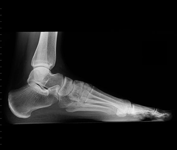
- X-Ray – X-rays may be taken from a side view and the top of the foot. These views allow the physician to locate the coalition as well as determine how much wear and tear has occurred in the joints.
- Computer Tomography (CT) Scan – A CT scan is the gold standard for diagnosing a tarsal coalition. This is because it is a special type of x-ray that uses a computer to create detailed images of the joints. A CT scan becomes a viable option when x-rays fail to show the location of the symptoms or fail to provide a clear view of the coalition.
- Magnetic Resonance Imaging (MRI) – MRI can also be used to visualize and diagnose a tarsal coalition. The technique is useful in identifying a coalition that is composed of tissues instead of bones e.g. nonosseous fibrous and cartilaginous coalitions.
Treatment Options
Nonsurgical treatment options are used to curb the symptoms of tarsal coalition and reduce the motion at the affected joint. The treatment options include physical therapy, oral medications, steroid injections, immobilization, orthotic devices and injection of an anesthetic agent.
Surgery is considered if nonsurgical treatment options fail. The procedure is performed to either remove the bar in an attempt to reinstate normal motion between the affected bones or fuse the affected joints together firmly.


